Be Ready.
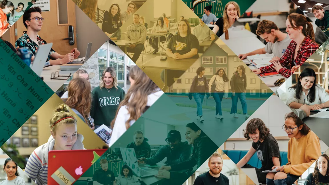
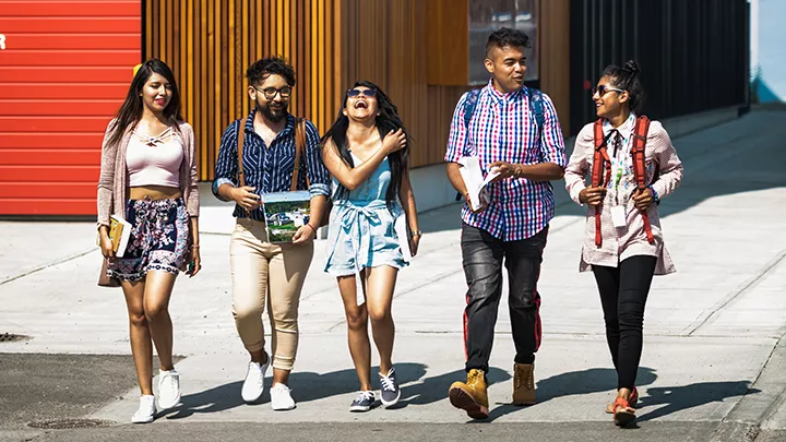
Be ready to elevate your potential, explore new opportunities, and impact your world. Find the program for you and be ready to shape your future.
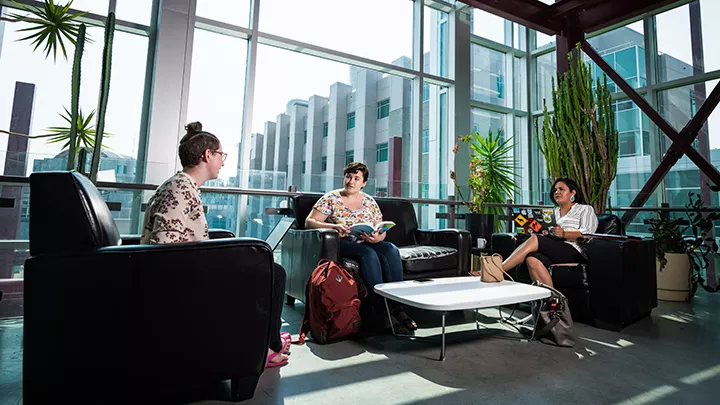
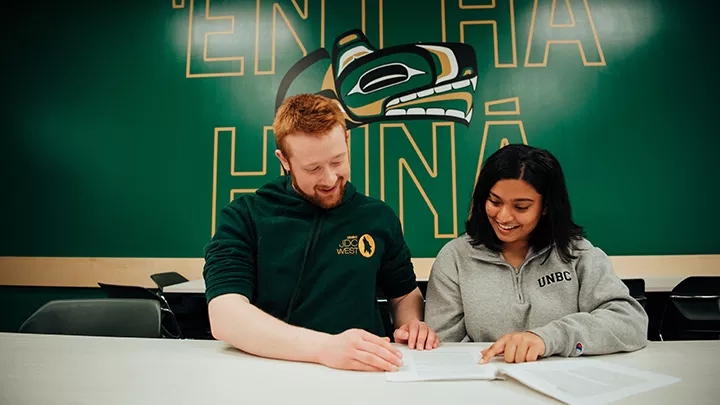
Join a community of enthusiastic researchers who will cultivate your curiosity for knowledge and exploration.
4
Canada Research Chairs
32
Graduate degree programs
We are on a path to meaningfully enact reconciliation with Indigenous Peoples, through dialogue, education, research, relationships, and service.
14%
of all students self-identify as Indigenous
50+
Reconciliation Initiatives
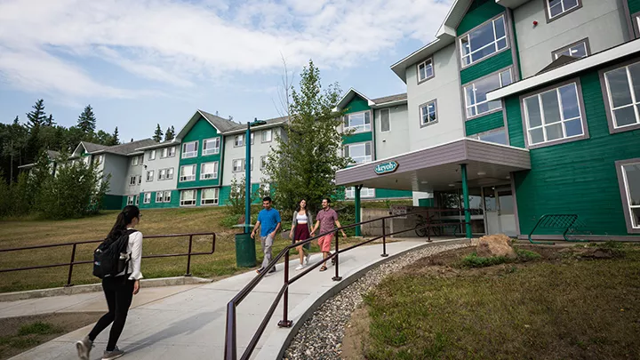
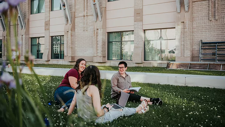
UNBC graduates are empowering northern communities as they put their degrees to work across the region and around the world.
16,000+
Alumni
81%
Graduates working in fields related to their programs
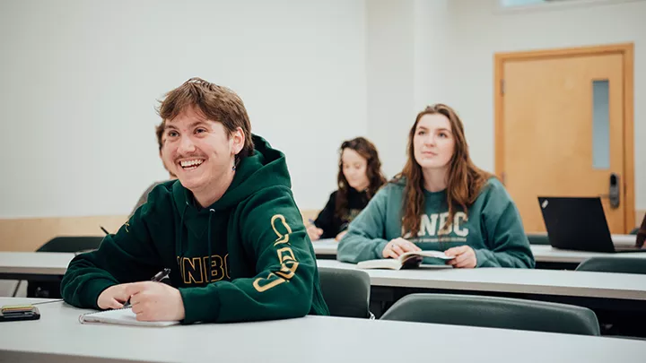
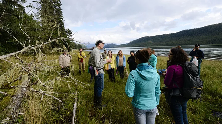
Apply the knowledge you learn in the classroom and discover solutions that can have a global impact.
17+
Programs with field school opportunities
36
Programs with co-op opportunities
UNBC consistently places as one of Canada’s top universities in national and international rankings.
#1
For students who win national awards
Top 5%
Of universities in the world
For thousands of years, Indigenous Peoples have walked gently on the diverse traditional territories where the University of Northern British Columbia community is grateful to live, work, learn, and play. We are committed to building and nurturing relationships with Indigenous Peoples, we acknowledge their traditional lands, and we thank them.
UNBC’s motto, from the Dakelh (Carrier) Elders, reminds us that all people have a voice and a viewpoint. Interpreted as "respecting all forms of life," 'En Cha Huná encapsulates the spirit of academic freedom, respect for others, and willingness to recognize different perspectives.